Microarraying
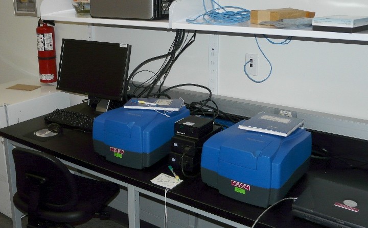
Axon GenePix 4000B Microarray Scanners

In order to visualize DNA microarrays hybridized to a probe of interest, the different dyes incorporated into the probes are stimulated to fluoresce by specific lasers which scan the microarray slide and produce a digital image. The CSFG currently possesses two GenePix 4000B Microarray Scanners which contain two different sets of lasers (Scanner #1 has a 532 nm and a 635 nm laser; Scanner #2 has a 473 nm and a 594 nm laser) which can activate the fluorescence of a wide variety of available dyes. The dyes emit different wavelengths of light at different intensities (depending on the degree of hybridization) and this light is passed through filters prior to being detected by photo-multiplier tubes. A sensitivity of as low as 0.1 fluors per square micrometer can be achieved. Since the dyes are stimulated by different lasers and emit different wavelengths of light, up to 4 different probes can be hybridized to the same array at the same time. The resolution of the images generated by the scanner ranges from 40 µm, for a quick preview scan, to 5 µm, for a high resolution data scan. At the highest resolution, a scan of the entire slide surface takes approximatley 12 minutes.
Location: GE S101
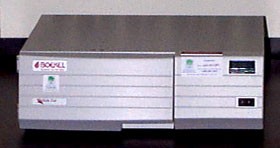
Boekel Inslide Out Hybridization Oven

The InSlide Out hybridization oven can be used for various in situ experiments but its primary use is for hybridizing fluorescently labelled DNA probes to microarrays. The oven can be set to temperatures ranging from about 30° - 70° C, with a +/- 0.2° C stability, and up to 18 slides can be processed at once. The tight seal of the hybridization tray prevents buffer evaporation.
Location: GE S101
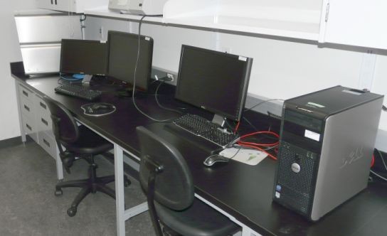
Imaging workstations

Computer workstations in this room are made available to users for data acquisition, for data analysis and for teaching-related purposes. In addition to the Leica Confocal Acquisition Software and the Genepix microarray data analysis software, a number of other software have been installed for data analysis.
Location: GE S101
