State-of-the-art microscopes
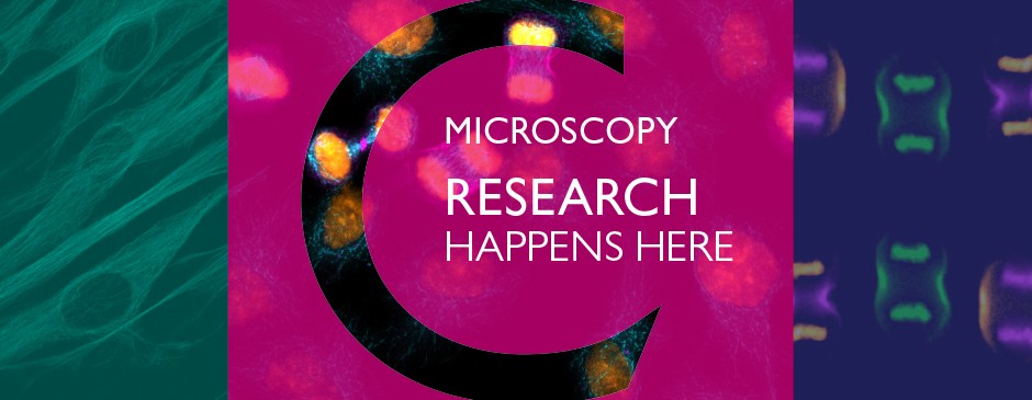
The Centre for Microscopy and Cellular Imaging provides members of Concordia University with access to advanced fluorescence microscopes.
Nikon Super-Res Spinning Disk
Zeiss Spinning Disk Confocal
Nikon Ti Epifluorescence Microscope
Nikon C2 Confocal/TIRF
Olympus Fluoview FV10i Laser scanning microscope
Arcturus XT Laser Capture Microdissection System
We have state-of-the-art microscopes customized for live cell imaging, with unique features like near-field, high-speed confocal at high magnification. On-site specialists will help you select the most suitable microscope and imaging technique for your project and provide training to lab personnel.
Cancer cell cytoskeleton
Rat Medium Spiny neuron from Dorsal Striatum
Golgi-labelled Hippocampal rat neurons
ECT2 in HeLa cell
Arabidopsis pavement cells (40x)
Human fibroblasts (Nikon C2 Confocal, Miroslav Milev)
Everything on how to use the CMCI from A to Z. First, you need to know how to prepare for your visit. Then read our requirements, rules, regulations, fees, booking and cancellation policies, acknowledgements, and instructions on becoming a new user.