Zeiss Axioplan Fluorescence Microscope

The Zeiss Axioplan fluorescence microscope is mounted with a Lumenera Infinity 3-1C 1.4 megapixel color cooled CCD camera and can be used to perform compound digital micrography at the CSFG. The microscope is equipped with 10x, 40x (DRY), 63x (OIL), 63x (OIL DIC) and 100x (OIL) objectives and other optic components required to perform brightfield, phase contrast and differential interference contrast microscopy. Illumination for epi-fluorescent excitation is provided an X-cite 120Q illuminator from EXFO.
Filter set A:
UV Filter Cube #487902 (Exciter Filter: G 365, Beam Splitter: FT 395, Barrier Filter: LP 420). This cube is used to visualize UV-excitable fluors like DAPI.
Blue Filter Cube #487910 (Exciter Filter: BP 450-490, Beam Splitter: FT 510, Barrier Filter: BP 515-565). Fluorescein, GFP and Oregon Green can be visualized using this filter cube.
Green Filter Cube #487915 (Exciter Filter: BP 546±12, Beam Splitter: FT 580,
Barrier Filter: LP 590). Green-excitable fluors like rhodamine and Cy3 can be visualized using this filter cube.
Filter set B:
GFP Filter Cube #1031346 (Exciter Filter: BP 470/40, Beam Splitter: FT 495, Barrier Filter: BP 525/30). This cube is used to visualize eGFP, CFP, GFP (S65T).
dsRed Filter Cube #1114462 (Exciter Filter: BP 560/40, Beam Splitter: FT 585, Barrier Filter: BP 630/75). This cube is used to visualize dsRed and mRFP.
YFP Filter Cube #1196681 (Exciter Filter: BP 500/20, Beam Splitter: FT 515,
Barrier Filter: BP 535/30). This cube is used to visualize eYFP, eGFP and Alexa 488.
Location: GE S105.
Imaging workstations

Computer workstations in this room are made available to users for data acquisition, for data analysis and for teaching-related purposes. In addition to the Leica Confocal Acquisition Software and the Genepix microarray data analysis software, a number of other software have been installed for data analysis.
Location: GE S101.
Singer MSM Micromanipulator

The micromanipulator is primarily used for spore dissection of sporulating yeasts. Using a microscope for visualization (4x and 20x objective lenses) and a highly precise movable stage (resolution of 4 µm and repeatability of 2 µm) and an extremely thin tapered glass capillary needle (40 - 50 µm diameter) for manipulation, spores can be separated and placed on pre-determined sections of a solid growth-medium plate. In such a manner, strains with different genotypes can be isolated.
Location: SP 444.01
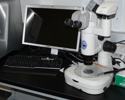
Nikon SMZ1500 Stereomicroscope

The Nikon SMZ1500 is a stereomicroscope with a 15:1 zoom ratio and a 0.75X to 11.25X zoom range. A 10X eyepiece allows total magnification of 7.5X to 112.5X. Illumination is provided by a 30W halogen lamp in the base and/or by an external 150W fiber optic episcopic illuminator that can provide bright illumination over the entire surface of the sample. A beam splitter allows images to be captured on a Leica DFC420 5 megapixel color digital camera.
Location: GE S105.
Nikon TMS Inverted Microscope

The Nikon TMS is an inverted microscope basic phase contrast capability (10x, 20x and 40x). Illumination is provided by a 20W halogen lamp in the top of the microscope. A beam splitter allows images to be captured on a SPOT Insight 2 megapixel color digital camera.
Location: GE S110-01. Access restricted to users of this facility.
